
a
b
c
e
g
h
i
j
l
m
n
p
s
v
x
a
anisotropicˌanʌɪsəˈtrəʊpɪk – adjective.
A material that has physical properties which have different magnitudes in different directions. Of a property: varying with direction. Opposite to isotropic. This is the property that makes some crystals display various colors when sandwiched between two polarizer filters.
anisotropically – adverb
anisotropy – noun
Ascorbic acid – Vitamin C
A natural water-soluble vitamin (Vitamin C), which can be used to make crystals for polarized micrograph. 1 gram dissolves in 3 ml of water. It will also dissolve in ethanol, so can use half water half ethanol for a quicker drying solution.
https://pubchem.ncbi.nlm.nih.gov/compound/54670067
b
birefringence – Means double refraction. This is the quality required in a subject such as crystals to make them colorful when sandwiched between two polarizers on a microscope.
birefringent adjective having double refraction
c
Canada balsam (Xylene)
Canada balsam is a commonly used mounting medium to prepare permanent slides for microscopy. It is produced from the resin of the balsam fir tree and can be combined with xylene-containing specimens. (Refractive index (20°C) 1.515 – 1.530).
Citric Acid
Anhydrous Citric Acid is freely soluble in Ethanol, and although can be mixed with 50% water and 50% IPA (Isopropyl alcohol) it takes much longer, sometimes days to dry and form crystals, and so is not recommended with IPA.
https://pubchem.ncbi.nlm.nih.gov/compound/311
e
Endodermis (Starch Sheath)
The endodermis (the innermost layer of the cortex adjacent to the pericycle) is composed of closely packed cells that have within their walls Casparian strips, water-impermeable deposits of suberin that regulate water and mineral uptake by the roots.
Here is a photo showing the Endodermis of an Azalea plant stem in bright red, stained with Safranin.
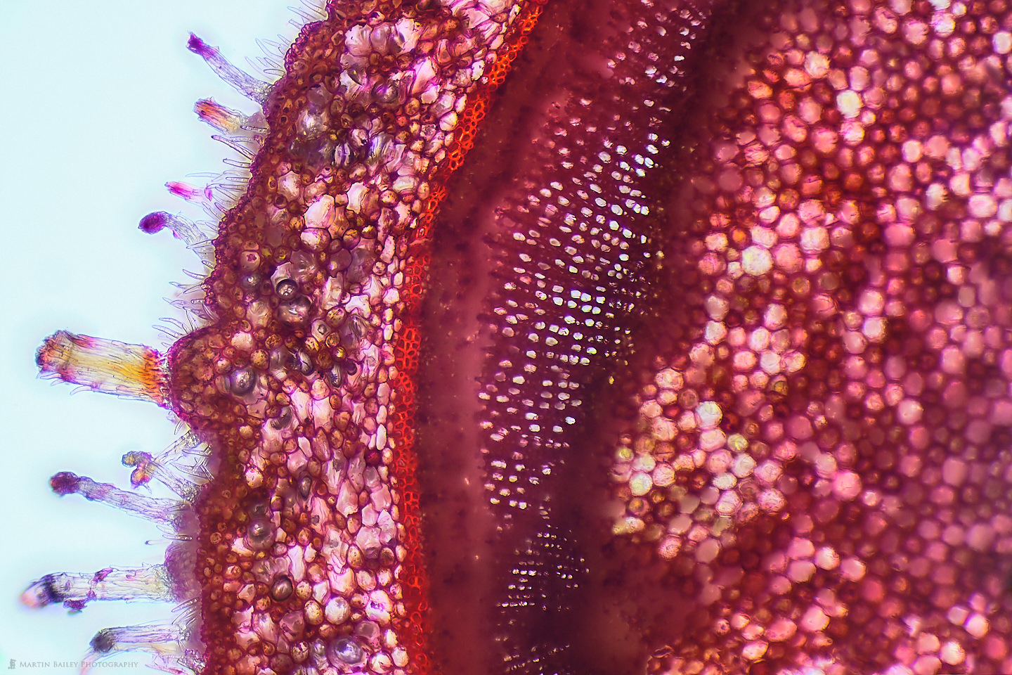
Azalea Stem (100X 64f Stack Safranine)
Eosin Y (stain)
Eosin Y Stain is a reversible, fluorescent red, acidic dye, commonly used in hospital histology labs. Eosin’s most important medical uses are in blood and bone-marrow testing, including the PAP smear. It can also test for protein in plant, animal and blood specimens. This stain is a valid substitute for Congo Red or Neutral Red and is frequently used as a counter-stain to Haematoxylin in H&E staining.
g
Gentian violet (stain)
Gentian Violet is a blue, aniline-derived dye with antifungal and antimitotic properties. Gentian violet (GV) dissociates into positive (GV+) and negative ions (Cl-) that penetrate both gram-positive and gram-negative bacterial cells. The GV+ ions interact with negatively charged components of the bacterial cell wall including lipopolysaccharide , peptidoglycan and DNA. This agent is also a mutagen and mitotic poison. GV elicits a photodynamic action mediated by a free-radical mechanism. Furthermore, this agent dissipates the action potential on prokaryotic or eukaryotic membranes by inducing permeability, thereby leading to respiratory inhibition and subsequent cell death.
h
Hematoxylin (stain)
Tissue stain that can be combined with Eosin.
i
Isopropyl alcohol (IPA)
Isopropyl Alcohol is an isomer of propyl alcohol with antibacterial properties. Although the exact mechanism of isopropanol’s disinfecting action is not known. Isopropanol is used in soaps and lotions as an antiseptic.
Can be used with some substances to aid in the forming of crystals, and also can be used for cleaning microscope slides etc.
https://pubchem.ncbi.nlm.nih.gov/compound/3776
j
Janus Green (stain)
Ready-to-use green stain for normalizing antibody signal intensity to total cell amounts during in-cell ELISA (ab111622).
Cells fixed to a solid surface (typically in microplate wells) can be stained by adding 1X Janus Green Stain. Incubate cells for 5 minutes at room temperature. Dye should be removed and washed 5 times in ultrapure water or until excess dye is removed. The last wash should be removed and add 0.1 mL 0.5 M HCl per well and incubate for 10 minutes. Shake the plate for 10 seconds, and measure the OD 595 nm using a standard microplate spectrophotometer.
l
Light Green (stain)
Used in the preparation of the staining solution which is widely used as a counterstain. It is the standard dye in North America for staining collagen and is also used extensively in plant histology. It has a role as a histological dye.
m
Methal green pyronin (stain)
The Methyl Green Pyronin (RNA DNA Stain) is intended for use in the histological visualization of DNA, RNA and mast cell granules
Methylene Blue (stain)
Methylene Blue is a popular alkaline stain used to view microscopic life in brilliant color. It helps make cells show up against their background, where their shape can help you determine what they are (their morphology).
It’s attraction to acid makes it particularly useful for viewing animal cells since these cell nuclei contain deoxyribonucleic acid (DNA). It’s also used in aquariums to guard against fungal infections in fish.
microscope ˈmʌɪkrəskəʊp noun & verb
An optical instrument, consisting of a lens or combination of lenses, which produces a magnified image of an object close to it so as to reveal details invisible to the naked eye.
Microtome mʌɪkrə(ʊ)təʊm nound & verb
An instrument for cutting extremely thin sections for microscopic work.
n
Neutral Red (stain)
The Neutral Red (Toluylene Red) Solution can be used as a red nuclear counterstain in various histological procedures. This solution has been proven to generate excellent nuclei staining on frozen tissue sections and paraffin-embedded brain sections with or without enzyme histochemistry and immunohistochemistry. The staining can be carried out at room temperature (18-25oC) and takes approximately 1-5 minutes. Staining time should be optimized for each tissue type and staining intensity desired. This counterstain solution is suitable for use with non-aqueous mountants.
p
plasmolysis plazˈmɒlɪsɪs noun
Contraction of the cytoplasm of a plant cell with separation of the plasma membrane from the cell wall, due to the osmotic withdrawal of liquid into a medium of high concentration.
s
Safranin (stain)
Safranin is a cationic dye used in histology and cytology to distinguish and identify different tissues and cells. It is popular in medical research for staining acidic proteoglycan that is found in cartilage tissues, enabling the researchers to analyze cell chondrogenesis. The safranin is employed as a counter-stain in endospore staining and Gram’s staining. It is mostly utilized for the identification of cartilage, mucin, and mast cell granules.
The safranin stain works by binding to acidic proteoglycans in cartilage tissues with a high affinity forming a reddish orange complex. The binding made cartilage tissues appear red when observed under the microscope. The safranin staining helps the researchers detect not only cartilage tissues but also all the body tissues and organs. The safranin stain is one of the most promising safe-lab stains used for histopathological and cytological research.
Staining procedures (for Safranin)
The following procedure should be followed for safranin staining.
To prepare the staining solution
- Add 20mg safranin powder to a 100ml beaker.
- Pour 20ml distilled water in the beaker and make 0.1% safranin staining solution by constant stirring.
- Transfer 20mg of fast green dye in another 100ml beaker. Moreover, make it 0.1% staining solution by adding 20ml distilled water in it.
- Filter both the staining solutions to avoid particles.
To prepare slides for observation
- Hydrate the slides after deparaffinization.
- After washing the slides, stain them with the fast green solution for 5-10 minutes.
- Rinse the slides with 0.1% acetic acid for 10-15 seconds.
- Stain the slides with 0.1% safranin solution for 20-30 minutes.
- Clear and dehydrate the slides (Dehydrate in 95% and 100% ethanol)
- Observe the slides under the microscope for qualitative and quantitative analysis.
Sodium sulfite – Na2SO3 or Na2O3S
Handling caution required.Can be used to make crystals on a slide for micrography.Soluble in water, but only sparingly soluble in alcohol.
Relatively safe to handle in dry powder form but toxic if ingested, and corrosive and irritant when wet.https://pubchem.ncbi.nlm.nih.gov/compound/Sodium-sulfite
Stain
There are various substances used to stain specimens to make them easier to see and photograph, or sometimes, just to make them look more beautiful! Jump to the following letters to see details of:
Eosin Y
Gentian violet
Hematoxylin
Janus Green
Light Green
Methal green pyronin
Methylene Blue
Neutral Red
v
Vitamin C
Can be used to make crystals for polarized micrograph – See “Ascorbic Acid”.
x
Xylene (Canada) balsam
Canada balsam is a commonly used mounting medium to prepare permanent slides for microscopy. It is produced from the resin of the balsam fir tree and can be combined with xylene-containing specimens. (Refractive index (20°C) 1.515 – 1.530).
